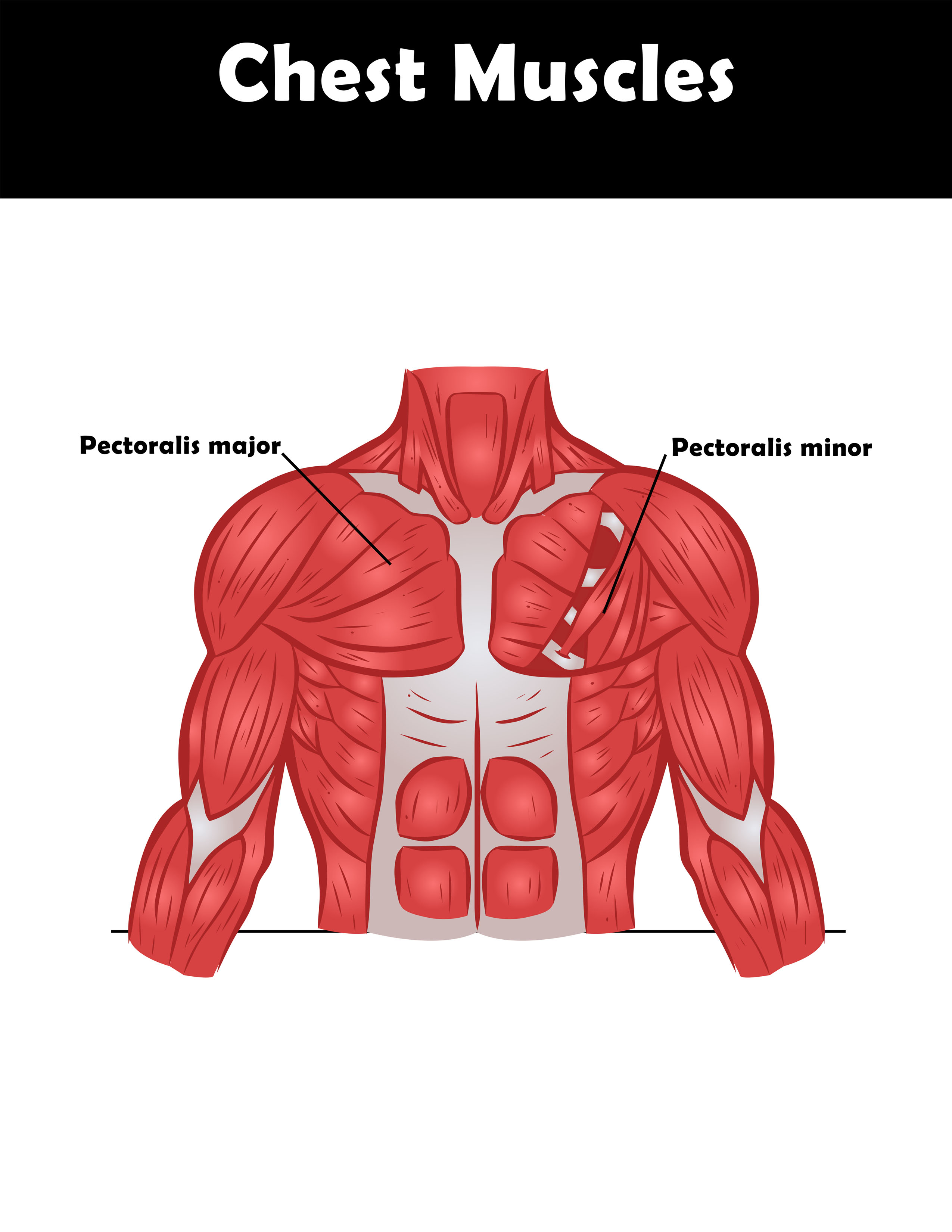
Labeled Chest Muscles Diagram / Just above the right hip) as shown on the diagram on the right.
byAdmin•
0
Labeled Chest Muscles Diagram / Just above the right hip) as shown on the diagram on the right.. The sensors can be placed on the forearms and leg as shown on the diagram on the left. Or they can be placed on the chest near the arms and above the right, lower abdomen (i.e. Spend some time revising this diagram by connecting the name and location of each structure with what you've just learned in the video. Just above the right hip) as shown on the diagram on the right. Dog anatomy is not very difficult to understand if a labeled diagram is present to provide a graphic illustration of the same.
The scapula is a wide, flat bone lying on the thoracic wall that provides an attachment for three groups of muscles: The ascending aorta is a portion of the aorta beginning at the upper part of the base of the left ventricle, on a level with the lower border of the third costal cartilage behind the left half of the sternum; Spend some time revising this diagram by connecting the name and location of each structure with what you've just learned in the video. It provides information about a dog's skeletal, reproductive, internal, and external anatomy, along with accompanying labeled diagrams. Just above the right hip) as shown on the diagram on the right.
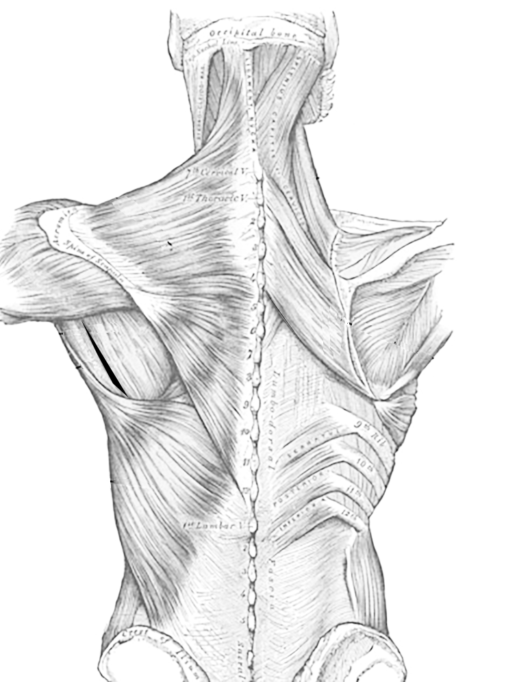
Muscles of the Thoracic Region, Dorsal Side from www.biologycorner.com Just above the right hip) as shown on the diagram on the right. Each arises from the lower border of a rib, and is inserted into the upper border of the rib below. It provides information about a dog's skeletal, reproductive, internal, and external anatomy, along with accompanying labeled diagrams. It passes diagonally upward, forward, and to the right, in the direction of the heart's axis, as high as the upper border of the second right costal cartilage. Take a look at the leg muscles diagram below, where you see each muscle clearly labeled. May 31, 2021 · leg muscles labeled. The internal intercostal muscles relax while the external muscles contract causing the expansion of the chest cavity and an influx of air into the lungs. Plasma red blood cell the region labeled x represents part of a) a glomerulus c) a villus b) an alveolus d) the liver 13.
Standard anatomical position in humans.
The diagram below represents part of a capillary in a specific region of the human body. The ascending aorta is a portion of the aorta beginning at the upper part of the base of the left ventricle, on a level with the lower border of the third costal cartilage behind the left half of the sternum; Spend some time revising this diagram by connecting the name and location of each structure with what you've just learned in the video. The scapula is a wide, flat bone lying on the thoracic wall that provides an attachment for three groups of muscles: Just above the right hip) as shown on the diagram on the right. These muscles work in unison when inhalation occurs. Intrinsic, extrinsic, and stabilising and rotating muscles. The sensors can be placed on the forearms and leg as shown on the diagram on the left. The aim of this exercise is to improve your confidence in identifying different structures. Take a look at the leg muscles diagram below, where you see each muscle clearly labeled. It passes diagonally upward, forward, and to the right, in the direction of the heart's axis, as high as the upper border of the second right costal cartilage. Dog anatomy is not very difficult to understand if a labeled diagram is present to provide a graphic illustration of the same. In this position, a person is standing upright with the lower limbs together or slightly apart, feet flat on the floor and facing forward, upper limbs at the sides with the palms facing forward and thumbs pointing away from the body, and head and eyes directed.
Plasma red blood cell the region labeled x represents part of a) a glomerulus c) a villus b) an alveolus d) the liver 13. Intrinsic, extrinsic, and stabilising and rotating muscles. It passes diagonally upward, forward, and to the right, in the direction of the heart's axis, as high as the upper border of the second right costal cartilage. Standard anatomical position in humans. Each arises from the lower border of a rib, and is inserted into the upper border of the rib below.
Lab 6: Axial Related Muscles (Trunk, Neck & Head ... from s3.amazonaws.com Take a look at the leg muscles diagram below, where you see each muscle clearly labeled. That is exactly what you will find in this dogappy article. The standard anatomical position is agreed upon by the international medical community. It passes diagonally upward, forward, and to the right, in the direction of the heart's axis, as high as the upper border of the second right costal cartilage. The scapula is a wide, flat bone lying on the thoracic wall that provides an attachment for three groups of muscles: These muscles work in unison when inhalation occurs. In this position, a person is standing upright with the lower limbs together or slightly apart, feet flat on the floor and facing forward, upper limbs at the sides with the palms facing forward and thumbs pointing away from the body, and head and eyes directed. The diagram below represents part of a capillary in a specific region of the human body.
Which structure shown in the diagram below contracts, causing a pressure change in the chest cavity during breathing?
The diagram below represents part of a capillary in a specific region of the human body. Just above the right hip) as shown on the diagram on the right. Dog anatomy is not very difficult to understand if a labeled diagram is present to provide a graphic illustration of the same. These muscles work in unison when inhalation occurs. The standard anatomical position is agreed upon by the international medical community. The intrinsic muscles of the scapula include the muscles of the rotator cuff—the subscapularis, teres minor, supraspinatus, and infraspinatus. Each arises from the lower border of a rib, and is inserted into the upper border of the rib below. Standard anatomical position in humans. Intrinsic, extrinsic, and stabilising and rotating muscles. The scapula is a wide, flat bone lying on the thoracic wall that provides an attachment for three groups of muscles: That is exactly what you will find in this dogappy article. It provides information about a dog's skeletal, reproductive, internal, and external anatomy, along with accompanying labeled diagrams. Program within @mayoclinicgradschool is currently accepting applications!
In this position, a person is standing upright with the lower limbs together or slightly apart, feet flat on the floor and facing forward, upper limbs at the sides with the palms facing forward and thumbs pointing away from the body, and head and eyes directed. It provides information about a dog's skeletal, reproductive, internal, and external anatomy, along with accompanying labeled diagrams. Standard anatomical position in humans. Take a look at the leg muscles diagram below, where you see each muscle clearly labeled. Dog anatomy is not very difficult to understand if a labeled diagram is present to provide a graphic illustration of the same.
Best Chest Exercises from www.makeoverfitness.com The scapula is a wide, flat bone lying on the thoracic wall that provides an attachment for three groups of muscles: The intrinsic muscles of the scapula include the muscles of the rotator cuff—the subscapularis, teres minor, supraspinatus, and infraspinatus. It passes diagonally upward, forward, and to the right, in the direction of the heart's axis, as high as the upper border of the second right costal cartilage. The internal intercostal muscles relax while the external muscles contract causing the expansion of the chest cavity and an influx of air into the lungs. The standard anatomical position is agreed upon by the international medical community. Just above the right hip) as shown on the diagram on the right. Each arises from the lower border of a rib, and is inserted into the upper border of the rib below. Jun 17, 2021 · heart muscles work constantly (thank goodness!), so the heart has a very high nutrient need.
Spend some time revising this diagram by connecting the name and location of each structure with what you've just learned in the video.
Take a look at the leg muscles diagram below, where you see each muscle clearly labeled. Program within @mayoclinicgradschool is currently accepting applications! It passes diagonally upward, forward, and to the right, in the direction of the heart's axis, as high as the upper border of the second right costal cartilage. In this position, a person is standing upright with the lower limbs together or slightly apart, feet flat on the floor and facing forward, upper limbs at the sides with the palms facing forward and thumbs pointing away from the body, and head and eyes directed. Jun 17, 2021 · heart muscles work constantly (thank goodness!), so the heart has a very high nutrient need. Intrinsic, extrinsic, and stabilising and rotating muscles. It provides information about a dog's skeletal, reproductive, internal, and external anatomy, along with accompanying labeled diagrams. That is exactly what you will find in this dogappy article. These muscles work in unison when inhalation occurs. The scapula is a wide, flat bone lying on the thoracic wall that provides an attachment for three groups of muscles: The sensors can be placed on the forearms and leg as shown on the diagram on the left. Just above the right hip) as shown on the diagram on the right. The internal intercostal muscles relax while the external muscles contract causing the expansion of the chest cavity and an influx of air into the lungs.
The aim of this exercise is to improve your confidence in identifying different structures chest muscles diagram. The ascending aorta is a portion of the aorta beginning at the upper part of the base of the left ventricle, on a level with the lower border of the third costal cartilage behind the left half of the sternum;